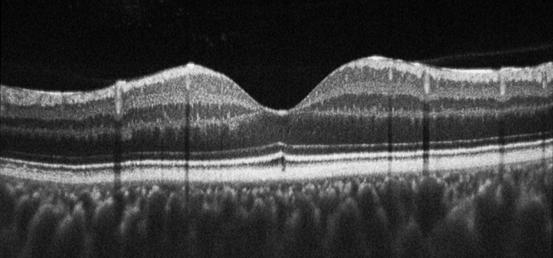

3– 7 For example, constant, intense light specifically results in the death of photoreceptor cells in the dorsal and central retina. 1, 2 Furthermore, the damaged zebrafish retina, unlike the mammalian retina, undergoes a spontaneous, robust, and specific regeneration response after many different insults. Zebrafish is a leading model system to study retinal development and degeneration. Thus, SD-OCT provides a noninvasive and quantitative method to assess the morphology and the extent of damage and repair in the zebrafish retina. SD-OCT was less accurate at detecting the inner nuclear layer in ouabain-damaged retinas, but accurately detected the undamaged outer nuclear layer. SD-OCT accurately represented retinal lamination and photoreceptor loss and recovery during light-induced damage and subsequent regeneration. Axial measurements of SD-OCT also revealed vitreal morphology that was not readily visualized by histology. Measurements between SD-OCT and histology were very similar for the undamaged, damaged, and regenerating retinas. Images were captured and the measurements of retinal morphology were made by SD-OCT, and then compared with those obtained by histology of the same eyes. Retinas of control dark-adapted adult albino zebrafish were compared with retinas subjected to 24 hours of constant intense light and recovered for up to 8 weeks or ouabain-damaged retinas that recovered for up to 3 weeks. Experiments assessed the ability of spectral-domain optical coherence tomography (SD-OCT) to accurately represent the structural organization of the adult zebrafish retina and reveal the dynamic morphologic changes during either light-induced damage and regeneration of photoreceptors or ouabain-induced inner retinal damage.


 0 kommentar(er)
